
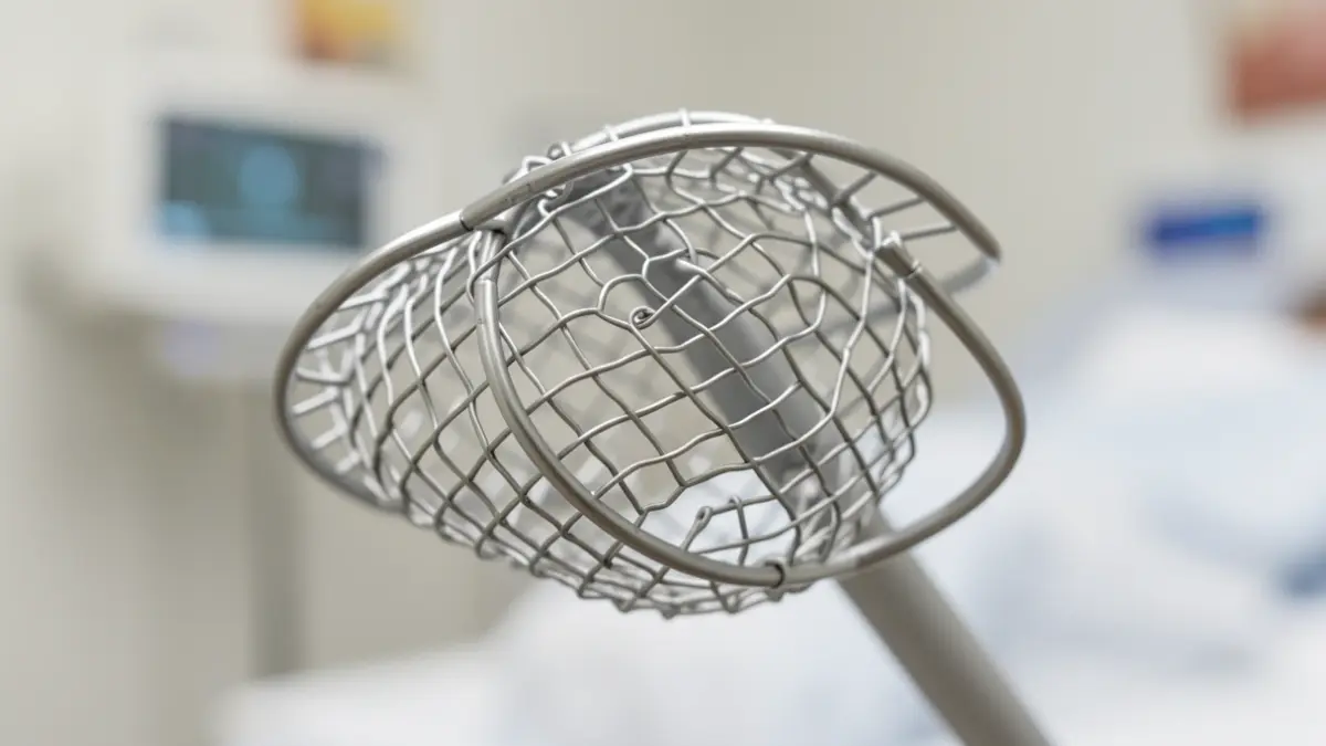
This advancement, resulting from collaboration between the Section of Vascular and Interventional Radiology, the Venous Thromboembolic Disease Unit of Internal Medicine, and the Hemostasis Section of Hematology and Hemotherapy, aims to increase care quality without raising associated complications. Inferior vena cava filters are devices implanted in the abdominal vena cava to intercept blood clots from the legs, acting as a 'safety net'.
According to Francisco Galeano, an internal medicine physician at the center, these devices are indicated in very specific situations, mainly for patients with recent deep vein thrombosis or pulmonary embolism who cannot receive anticoagulant treatment. However, their use in clinical practice has traditionally been heterogeneous, with off-guideline indications and low removal rates.
The new protocol establishes clear criteria for indication, follow-up, and removal, including periodic reviews to assess necessity and determine the optimal time for extraction, thereby ensuring maximum patient safety. The research, published in doi.org/10.1007/s11739-026-04361-7, compared data from 234 patients before and after implementation.
Results show a significant improvement in the appropriateness of filter indication (from 74.7% to 88.5%) and a notable increase in removal rates (from 41.2% to 65.4% at 90 days and from 41.8% to 73.1% at 180 days). Prolonged presence of these devices is associated with a higher risk of complications, such as new clot formation or altered blood flow.
The study confirms that the increase in removal rates has not led to a rise in adverse events. Furthermore, filter removal was independently associated with lower mortality at 180 days, reinforcing the importance of optimizing clinical management. The protocol has been reviewed by the Preventive Medicine Service and approved by the Venous Thromboembolic Disease Unit, with the support of the Complutense University of Madrid.